Posts

Showing posts from August, 2020

DNA REPLICATION PDF

Image
  DNA REPLICATION PDF  👈 click on this link to view and download PDF 

DNA MODEL OF SHASHISEKHRAN

Image
  शशिशेखरन का डीएनए मॉडल   👈 पीडीएफ देखने और डाउनलोड करने के लिए यहां क्लिक करें। पर मेरी वेबसाइट क्लिक पालन करने के लिए "दृश्य वेब संस्करण" 👇

Chargaff 's rules PDF

  Chargaff's rules PDF  👈 click on this link to view and download PDF

Chargaff's rules

  Chargaff's rules:- One other key piece of information related to the structure of DNA came from Austrian biochemist Erwin Chargaff. Chargaff analysed the DNA of different species, determining its composition of A, T, C, and G bases. He made several key observations:  A, T, C, and G were not found in equal quantities (as some models at the time would have predicted)  The amounts of the bases varied among species, but not between individuals of the same species  The amount of A always equalled the amount of T, and the amount of C always equalled the amount of G (A = T and G = C)  - Purines always equal to pyrimidine . ( A+G = T+C ) - MOLAR RATIO BETWEEN PURINE AND PYRIMIDINE IS ALWAYS 1 (1:1) -  BASE RATIO IN DNA ( A+T/G+C ) IS ALWAYS FIXED IN A SPECIES BUT DIFFERENT IN DIFFERENT SPECIES . In Prokaryotes = < 1 ( less than 1 )  In Eukaryotes = > 1 ( more than 1 )  These findings, called Chargaff's rules, turned out to be crucial to Watson a...
WATSON AND CRICK MODEL PDF

  WATSON AND CRICK MODEL PDF   👈 click on this link to view and  download PDF

WATSON AND CRICK MODEL OF DNA

Image
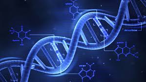
Watson and  Crick Model of DNA- - Two British scientist James D. Watson  and F.H.C. Crick(1953) presented a  universally accepted model of DNA . That  time they were working in Cambridge  University. According to them the DNA molecule  have high molecular weight and double  strands . - That time Watson and Crick also had  results of X-ray Diffraction collected by  Maurice Wilkins, Rosalind Franklin and his colleagues. Following are the main points of this  model- 1. Each DNA molecule is made up of two  polydeoxyribonucleotide strands. 2. Each polynucleotide strand is made up  of many nucleotides joined together by  phosphodiester bonds. 3. All N-base in nucleotides are attached  to the first carbon of deoxyribose sugar. 4. Thymine is located in second strand just  in front of the Adenine located in first  strand, similarly Cytosin is located in front of Guanine. These strand are called  Complementary S...

ASSEMBLY OF DIFFERENT COMPOUNDS IN DNA PDF

  ASSEMBLY OF DIFFERENT COMPOUNDS IN DNA  👈 click on this link to open pdf

ASSEMBLY OF DIFFERENT COMPOUNDS IN DNA

Image
  Assembly of different compounds in DNA:- Generally following three terms are used  to clearly understand the combination of  different chemical components in DNA- 1. Nucleoside 2. Nucleotide 3. Polynucleotide 1. Nucleoside- A molecule made from combination of  Nitrogen base and Pentose sugar is called  Nucleoside. Types of Nucleotides- A. Deoxyadenosine B. Deoxyguanosine C. Deoxythymidine D. Deoxycytidine 2. Nucleotide:- A molecule made from the combination of  a Nucleoside and phosphoric acid is called  Nucleotide. Nucleoside (Deoxyribose sugar + nitrogen  base) + Phosphoric acid =Deoxyribonucleotide Types of Nucleotide- A. Deoxyadenylic acid B. Dexyoguanylic acid C. Deoxycytidylic acid  D. Deoxythymidylic acid 3. Polynucleotide- Many Nucleotide monomers combine to  form a polynucleotide chain by  phosphodiester bonds.

CHEMICAL ORGANISATION OF DNA PDF

  CHEMICAL ORGANISATION OF DNA  👈 click on this link to view and download PDF

TYPES OF DNA PDF

  TYPES OF DNA  👈 click on this link to view PDF 

DNA INTRODUCTION PDF

  DNA INTRODUCTION PDF  👈 click on this link to view pdf

Chemical Organization Of DNA

Image
 Chemical organisation of DNA: - According to chemical analysis DNA is  produced by following three types of  compounds- 1. Pentose Sugar 2. Phosphate 3. Nitrogenous base 1.Pentose Sugar:- (A) It is 5 member ring. (B) 4 Carbon and 1 oxygen atoms form ring,  the 5th carbon atom is attached with the  group –CH2 on outside of the ring. (C) Deoxyribose sugar has one oxygen atom  absent at carbon No.2 compared to ribose  sugar. 2.Phosphate:- The phosphate group in DNA is present as  phosphoric acid. 3. Nitrogenous base: - On the chemical basis there is two types of  nitrogen bases- (a) Pyrimidine (b) Purines (a) Pyrimidine :- - These are nitrogen compounds of single  ring. - Its 6 member benzene ring have Nitrogen  atom at the site number 1 and 3. - A DNA molecule have two types of  pyrimidines- 1. Cytosine (2-Hydroxy-6-amino pyrimidine) 2. Thymine (2,6 Dihydroxy-5-methyle  pyrimidine) * In RNA uracil is found in place of thymi...

DNA TYPES

Image
  Types of DNA:- Following are the 7 main types of DNA- 1. The B-Form of DNA (B- DNA): Structure of B-form of DNA has been  proposed by Watson and Crick. It is  present in every cell at a very high relative  humidity (92%) and low concentration of  ions. It has antiparallel double helix,  rotating clockwise (right hand) and made  up of sugar- phosphate back bone  combined with base pairs or purine- pyrimidine. The base pairs are perpendicular to  longitudinal axis of the helix. The base  pairs tilt to helix by 6.3°. The B-form of  DNA is metabolically stable. The length of  a complete turn is 34 Angstroms and  number of bases pairs in a complete turn  is 10 (10.4). The diameter of helix is 20 Angstroms and height of each base pair is  3.38 Angstroms. Each base pair accounts  for 36(34.6 angstrom according to 10.4  base pair per helix turn, 360/10.4 =34.6) degree of rotation, creating a minor  ...

Preserve Biodiversity

Preserve Biodiversity  👈click on preserve Biodiversity

DNA

Image
INTRODUCTION  :- Full form – Deoxyribonucleic Acid First time Frederic Miescher discovered DNA in  pus cells in 1869 and called it nuclein. Because of Nuclein’s acidic nature in 1899  Richard Altman called it Nucleic Acid. History of Nucleic Acid 1. Freidrich  Miescher (1869) He discovered DNA in pus cells and  called Nuclein. 2. Richard Altman (1899 ) He called nuclein as Nucleic Acid. 3. O.F. Griffith (1928) He discovered Transformatin in  bacteria. 4. J.D. Watson and  F.H.C. Crick (1953) Described Double Helix Model of DNA. 5. Kuhn (1957 ) Calculated molecular weight of DNA. 6. H.F. Conart (1957 D) RNA in T.M.V. as genetic material  7. Meselson and  Stahl 1958 Described Semiconservative method  of DNA replication. DNA Nature:- Most organisms, plants, bacteria, animal  virus, bacteriophage and plant virus have  double strand DNA but some bacterial virus  such as phi*174 and S13 have single strand  DNA. Sh...